Whole-brain spectroscopic MRI of Glutamate Stress Response in Major Depressive Disorder (MDD) patients to Quantify the Behavioral Effects of Ketamine
Major depressive disorder (MDD) is a leading cause of disability worldwide, and most of the affected population will never receive diagnosis or treatment. Depression is also a major problem in brain tumor patients as it is a major side effect of radiation therapy; between 35-50% of glioblastoma patients suffer from depression. All currently available antidepressants act by the same mechanism (blocking the monoamine reuptake pumps for serotonin, norepinephrine, or dopamine) with limited efficacy. About one-third of patients fail to respond, and in responders the onset of action is slow (4-6 weeks). Even among responders, significantly depressed patients are prone to drug-resistance over time. This study proposes to study the effects of ketamine in the depressed population. Ketamine acts on the glutamatergic receptor as a non-selective antagonist, and in responders, it is effective within 24 hours.
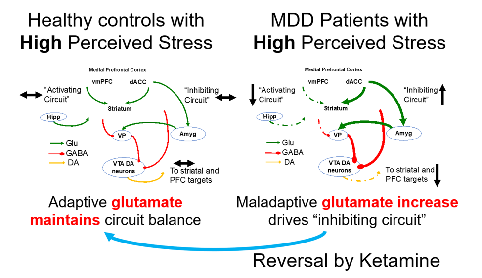
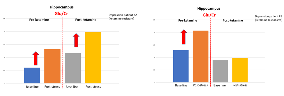
Spectroscopic MRI (sMRI) can be used for metabolite quantification, and our whole-brain imaging technique maps glutamate, glutamine, and glx concentrations within the brain to visualize the effects of ketamine treatment in MDD patients and matched controls. We hypothesize that healthy controls with increased chronic stress will demonstrate a decreased glutamate response to an acute stress challenge while MIDD patients with increased chronic stress will exhibit a relative increase of glutamate to acute stress. Preliminary results between a ketamine responsive and non-responsive MDD patient have shown promising outcomes.